To understand how cells work, it helps to be able to see their components clearly.
"If scientists can understand how cells work, then they can potentially understand disease states better, which might lead to better medicines down the road," Winter said. "If they can understand how plants are organized, they might be able to figure out how to take them apart to better utilize their components or turn them into biofuel."
But when it comes to visualizing the very tiny parts inside cells, even microscopes have their limit.
Arthur Winter, associate professor of chemistry, is expanding that limit by developing probes that enhance and simplify a relatively new microscopy technique called STORM (Stochastic Optical Reconstruction Microscopy) that lets scientists distinguish orders of magnitude smaller objects than they can with a typical microscope.
But when objects are smaller than approximately the wavelength of light the objects marked with dye stop being distinguishable from each other – this is known as the diffraction limit of light.
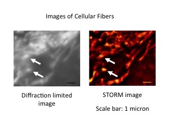
"Imagine that you are trying to resolve a meshwork of really small fibers," Winter said. "On the conventional microscope these just appear as a big blur."
STORM allows these small structures to be clearly measured. The "ST" in STORM stands for stochastic, which means random, and in this new approach objects are hit with light and randomly become visible. When the experiment is done correctly the odds are very low that two dye markers near each other will be visible at the same time.
This is important because when the fluorescent markers turn on at different times, they can be distinguished from what’s around them. In a STORM experiment, a series of images is collected that reveals flashes of light in different spots at different times, which can then be reconstructed into a single image that has much better resolution than would otherwise be possible.
Smith and Chamari Wijesooriya, a graduate student in chemistry, have built a STORM microscope here at Iowa State and collaborate with Winter as he develops chemical probes that will improve the technique, making it even more simple and effective for scientists.
“Fibrous bundles, cellulose, cell membranes — these are all structures that are smaller than the diffraction limit,” Winter said. “If you actually want to see how they’re embedded within the cell and how they’re organized, you really want to use a technique like this to be able to look at them."
The fluorescent markers that have been used in STORM techniques so far have their own drawbacks — once fluorescent, they often need a different wavelength of light to turn off this fluorescence so they don’t obscure the new dyes as they light up. The light source they use is also typically ultraviolet rather than visible light, and other additives are often needed to make the markers work as needed — but both ultraviolet light and additives can be damaging to cells and tissues.
Winter and Julie Peterson, a graduate student in chemistry, are creating new fluorescent markers that solve these issues. He takes advantage of a bleaching reaction to turn the fluorescence off with the same light source as it is exposed over a longer time.
"Everything eventually bleaches which is a side reaction that destroys the fluorophore and then switches it back off," Winter said.
The method developed by Winter, Smith and their students uses visible light and no additives, both of which are healthier for cells.
“The instrument is simpler, and the low laser power is less damaging to the sample,” Smith said. “This may make it possible to study many samples that are easily damaged by light with STORM imaging using simple instrumentation.”